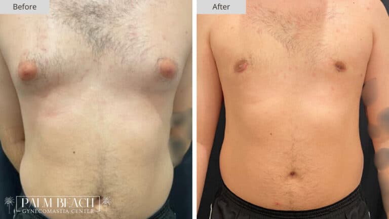
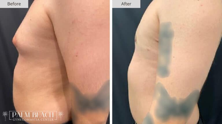
This male patient presented with one of the more pronounced cases of gland-dominant gynecomastia. From the side, the projection of breast tissue was clearly visible, forming a rounded bulge that sat noticeably forward on the chest wall. While his overall frame was athletic, the prominence of the chest tissue significantly altered his proportions and figure.
There was no visible sagging or skin excess, but the density and shape of the tissue strongly suggested that the issue was glandular rather than fatty. Palpation during physical exam confirmed a firm, rubbery mass directly beneath the nipple, extending into the surrounding subareolar area. The volume and consistency of the tissue meant that liposuction alone would have been ineffective.
This was a classic case where precision glandular excision would be essential.
Surgical Approach and Rationale
Our goal for this case was to restore a flat, masculine contour without creating irregularities, skin laxity, or visible scarring.
Based on the location and density of the tissue, we elected to perform direct gland excision through a periareolar approach. This method involves:
- A small semicircular incision made at the lower border of the areola
- Dissection to carefully isolate and remove the glandular mass
- Conservative sculpting to avoid contour depressions or tethering
- Layered closure to ensure optimal healing and minimal scar visibility
In cases of high projection, such as this one, there is a risk of over-resection. Care must be taken to avoid leaving a concavity behind, particularly when removing dense glandular tissue.
Our technique focused on balanced removal, keeping a thin layer of tissue behind the areola to maintain shape and avoid retraction.
Postoperative Healing and Recovery
The patient recovered smoothly. Mild swelling was present in the first week, along with expected tightness in the chest. He wore a compression garment full-time during this phase to help reduce inflammation and guide skin retraction.
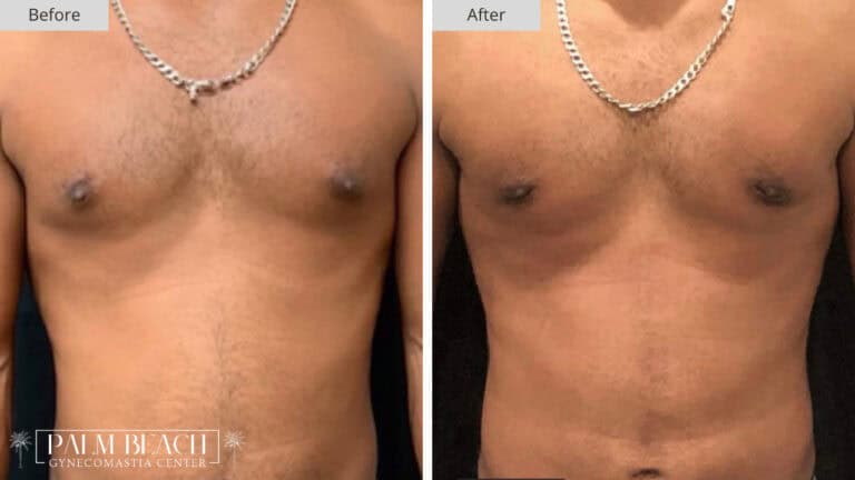
By the third week, the swelling had decreased, and the dramatic improvement in contour was clearly visible. The chest no longer projected outward. The area beneath the nipple lay flat against the chest wall, creating a more natural, athletic profile from the side.
There were no signs of fluid collection or asymmetry. The incision line had already begun to fade into the natural pigmentation of the areola.
Outcome at Six Weeks
By the six-week mark, the patient had:
- Full resolution of the visible chest projection
- Smooth, even contour across both sides of the chest
- No signs of puckering, tethering, or scar distortion
- High satisfaction with the shape, symmetry, and appearance in both relaxed and flexed positions
What made this case particularly impactful was the degree of transformation. From a side view, the bulge that had once defined his upper torso had been replaced with a clean, streamlined chest wall. The difference in posture, clothing fit, and self-perception was significant.
Surgeon’s Reflection
This case reinforces a key principle in gynecomastia surgery that not all chest fullness is the same, and not all patients need liposuction. In fact, for patients with firm, discrete glandular tissue and little surrounding fat, liposuction can be ineffective or even counterproductive if performed alone.
By tailoring our approach to the exact source of the problem, we achieved a dramatic and natural-looking result with minimal downtime and no visible signs of surgery. The patient now has the chest contour he always wanted, free from the burden of projection and self-consciousness.
At the Palm Beach Gynecomastia Center, this level of surgical customization is our standard. Whether the case is subtle or pronounced, our focus is always on delivering results that restore balance, confidence, and long-term satisfaction.